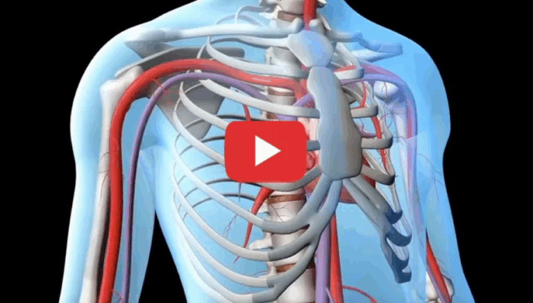
Intravascular Ultrasound (or IVUS) allows us to see a coronary artery from the inside-out. This unique point-of-view picture, generated in real time, yields information that goes beyond what is possible with routine imaging methods, such as coronary angiography, performed in the cath lab, or even non-invasive Multislice CT scans.
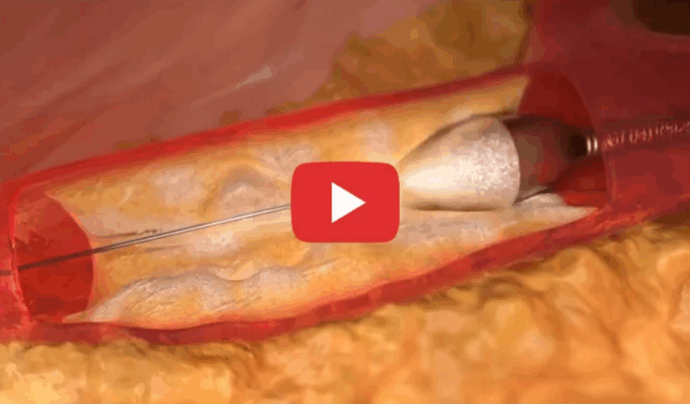
This cross-section view can aid in stent sizing, and in confirmation that the stent has been placed optimally, is fully expanded and hugging the vessel wall. A growing number of cardiologists feel that the new information yielded by IVUS can make a significant difference in how a patient is treated, and can provide for more accurate stent placement, reducing complications and the incidence of stent thrombosis.
How Does IVUS Work?
IVUS uses echocardiography: the same technology as the ultrasound imaging used in treadmill tests and many other medical exams. Very high frequency sound waves, called ultrasound, are emitted by a transducer. These ultrasound waves, which are beyond the range of human hearing, bounce off the various types of tissue structures in the body and the echo of these waves is then converted into a picture.
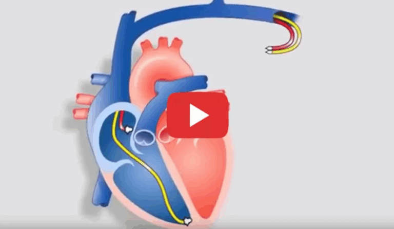
In the case of Intravascular Ultrasound, the transducers have been miniaturized to less than four hundredths of an inch and placed on the tip of a catheter. This catheter can be slipped into the coronary arteries over the same guide wire that is used to position angioplasty balloons or stents. It becomes, in effect, a tiny camera that gives us a cross-sectional view of the artery, a view that shows distinct circular layers, using shades of gray or colors.
OCT
In cardiology, OCT is an optical analog of intravascular ultrasound (IVUS), used to examine the coronary arteries. Ultrasound employed for IVUS examination is replaced by near-infrared light with a wavelength of about 1,300 nm, which is absorbed by red blood cells, water, lipids, and protein at relatively low levels. Through a rotating glass fiber-optic system, coherent infrared light can be directed and reflected within the tissue to create a detailed tissue image with extraordinarily high resolution, and IVUS-like cross-sectional tomographic images can be obtained. OCT and IVUS differ in several respects. The resolution of OCT (to 10-20 µm) is about 10-fold higher than that of IVUS (to 100-150 µm), but the maximum depth of tissue penetration is lower with OCT (1-2 mm) than with IVUS (4-8 mm). Another important difference is related to the strong attenuation of light by blood, which originates from two sources: absorption by hemoglobin and scattering by red blood cells. To examine coronary arteries, blood must first be removed during an OCT examination to eliminate massive scattering of light by red blood cells.