MBBS, MD’Clinical Ordinatura (Cardiology)
FACC, FICC, Fellowship in Intervention Cardiology
Adult and Paediatric Intervention Cardiologist

CALL FOR AN APPOINTMENT

HEART RHYTHM DISORDERS (ARRHYTHMIAS)

Heart Rhythm Disorders

Your heart is hard at work as it pumps blood and nutrients throughout your body. You can sometimes hear it or even feel it as it beats at a steady pace. It’s got an even, reliable rhythm that’s controlled by your body’s own electrical system. When that system has issues, though, you get a change in your heart’s rhythm that’s called arrhythmia.If you have an arrhythmia, it doesn’t necessarily mean you’ve got heart disease. There are many things that can cause your heart to flutter.

arrhythmias

What Causes Your Heart to Break Its Rhythm?

It’s possible to have a random arrhythmia even if your heart is healthy. If you do, talk to your doctor. Arrhythmias are caused by:

  • Infection or fever
  • Physical or emotional stress
  • Diseases such as anemia or thyroid disease
  • Drugs and other stimulants, such as caffeine, tobacco, alcohol, cocaine, amphetaminest and certain over-the-counter and prescription medications
  • Your genes
  • Some heart conditions
  • Symptoms of a Heart Rhythm Disorder

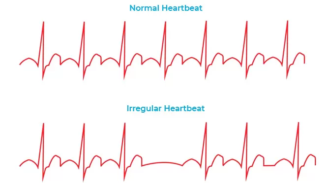
A typical heart will beat at 60 to 100 times per minute. It can beat faster if you need it to during exercise or in a stressful situation. It can slow down while you sleep. Your heart is used to slowing down and speeding up. That’s normal.

Noticeable Arrhythmia Symptoms May Include:

  • A fluttering in your chest
  • A racing heartbeat (tachycardia)
  • A slow heartbeat (bradycardia)
  • Chest pain
  • Shortness of breath
  • Lightheadedness or dizziness
  • Sweating
  • Fainting (syncope) or near fainting

What's Controlling Your Heartbeat?

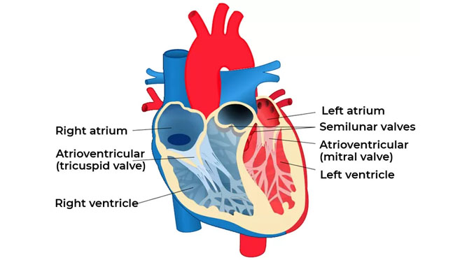
There’s a node in the upper right section of your heart that monitors your body’s need for blood. It’s called the sinoatrial (SA) or sinus node, and it acts like a natural pacemaker. It’s the main control and source of each heartbeat. It can speed up your heart rate when you need it, like when you exercise or get sick, or even when you feel happy. Your SA node sends out electrical impulses across the heart. These cause the chambers to contract at specific times, causing a heartbeat.

Types of Arrhythmias

Doctors classify arrhythmias not only by where they originate (atria or ventricles) but also by the speed of heart rate they cause:

  • Tachycardia (tak-ih-KAHR-dee-uh). This refers to a fast heartbeat – a resting heart rate greater than 100 beats a minute.
  • Bradycardia (brad-e-KAHR-dee-uh). This refers to a slow heartbeat – a resting heart rate less than 60 beats a minute.

Not all tachycardias or bradycardias mean you have heart disease. For example, during exercise it’s normal to develop a fast heartbeat as the heart speeds up to provide your tissues with more oxygen-rich blood. During sleep or times of deep relaxation, it’s not unusual for the heartbeat to be slower.

Tachycardias in the atria

Tachycardias originating in the atria include:

  • Atrial fibrillation. Atrial fibrillation is a rapid heart rate caused by chaotic electrical impulses in the atria. These signals result in rapid, uncoordinated, weak contractions of the atria 

The chaotic electrical signals bombard the AV node, usually resulting in an irregular, rapid rhythm of the ventricles. Atrial fibrillation may be temporary, but some episodes won’t end unless treated.

Atrial fibrillation may lead to serious complications such as stroke.

  • Atrial flutter. Atrial flutter is similar to atrial fibrillation. The heartbeats in atrial flutter are more-organized and more-rhythmic electrical impulses than in atrial fibrillation. Atrial flutter may also lead to serious complications such as stroke.
  • Supraventricular tachycardia. Supraventricular tachycardia is a broad term that includes many forms of arrhythmia originating above the ventricles (supraventricular) in the atria or AV node.
  • Wolff-Parkinson-White syndrome. In Wolff-Parkinson-White syndrome, a type of supraventricular tachycardia, there is an extra electrical pathway between the atria and the ventricles, which is present at birth. However, you may not experience symptoms until you’re an adult. This pathway may allow electrical signals to pass between the atria and the ventricles without passing through the AV node, leading to short circuits and rapid heartbeats.

Tachycardias in the ventricles

Tachycardias occurring in the ventricles include:

  • Ventricular tachycardia. Ventricular tachycardia is a rapid, regular heart rate that originates with abnormal electrical signals in the ventricles. The rapid heart rate doesn’t allow the ventricles to fill and contract efficiently to pump enough blood to the body. Ventricular tachycardia can often be a medical emergency. Without prompt medical treatment, ventricular tachycardia may worsen into ventricular fibrillation.
  • Ventricular fibrillation. Ventricular fibrillation occurs when rapid, chaotic electrical impulses cause the ventricles to quiver ineffectively instead of pumping necessary blood to the body. This serious problem is fatal if the heart isn’t restored to a normal rhythm within minutes.


Most people who experience ventricular fibrillation have an underlying heart disease or have experienced serious trauma, such as being struck by lightning.

  • Long QT syndrome. Long QT syndrome is a heart disorder that carries an increased risk of fast, chaotic heartbeats. The rapid heartbeats, caused by changes in the electrical system of your heart, may lead to fainting, and can be life-threatening. In some cases, your heart’s rhythm may be so erratic that it can cause sudden death.


You can be born with a genetic mutation that puts you at risk of long QT syndrome. In addition, several medications may cause long QT syndrome. Some medical conditions, such as congenital heart defects, may also cause long QT syndrome.

Bradycardia -A slow heartbeat

  • Although a heart rate below 60 beats a minute while at rest is considered bradycardia, a low resting heart rate doesn’t always signal a problem. If you’re physically fit, you may have an efficient heart capable of pumping an adequate supply of blood with fewer than 60 beats a minute at rest.

In addition, certain medications used to treat other conditions, such as high blood pressure, may lower your heart rate. However, if you have a slow heart rate and your heart isn’t pumping enough blood, you may have one of several bradycardias, including:

  • Sick sinus syndrome. If your sinus node, which is responsible for setting the pace of your heart, isn’t sending impulses properly, your heart rate may be too slow (bradycardia), or it may speed up (tachycardia) and slow down intermittently. Sick sinus syndrome can also be caused by scarring near the sinus node that’s slowing, disrupting or blocking the travel of impulses.
  • Conduction block. A block of your heart’s electrical pathways can occur in or near the AV node, which lies on the pathway between your atria and your ventricles. A block can also occur along other pathways to each ventricle.


Depending on the location and type of block, the impulses between the upper and lower halves of your heart may be slowed or blocked. If the signal is completely blocked, certain cells in the AV node or ventricles can make a steady, although usually slower, heartbeat.

Some blocks may cause no signs or symptoms, and others may cause skipped beats or bradycardia.

When should you reach out to us to get Medical Care

You may have noticed your heart racing, a fluttering in your chest, or a sensation that your heart skipped a beat. If this happens once or infrequently with no other symptoms; it’s usually not serious. Talk to us about your questions and concerns. If you get treatment and it doesn’t help, make sure to let us know as soon as you can. If you have any of the following symptoms, call us at +91 9591991809 right away:

  • Unexplained shortness of breath
  • Light-headedness or feeling faint
  • You feel that your heart is beating too slowly or too quickly
  • Chest pain
  • Chest pain with any of these symptoms

Diagnosis

To diagnose a heart arrhythmia, your doctor will review your symptoms and your medical history and conduct a physical examination. Your doctor may ask about – or test for – conditions that may trigger your arrhythmia, such as heart disease or a problem with your thyroid gland. Your doctor may also perform heart-monitoring tests specific to arrhythmias. These may include:

  • Electrocardiogram (ECG). During an ECG, sensors (electrodes) that can detect the electrical activity of your heart are attached to your chest and sometimes to your limbs. An ECG measures the timing and duration of each electrical phase in your heartbeat.
  • Holter monitor. This portable ECG device can be worn for a day or more to record your heart’s activity as you go about your routine.
  • Event monitor. For sporadic arrhythmias, you keep this portable ECG device available, attaching it to your body and pressing a button when you have symptoms. This lets your doctor check your heart rhythm at the time of your symptoms.
  • Echocardiogram. In this noninvasive test, a hand-held device (transducer) placed on your chest uses sound waves to produce images of your heart’s size, structure and motion.
  • Implantable loop recorder. This device detects abnormal heart rhythms and is implanted under the skin in the chest area. If your doctor doesn’t find an arrhythmia during those tests, he or she may try to trigger your arrhythmia with other tests, which may include:
  • Stress test. Some arrhythmias are triggered or worsened by exercise. During a stress test, you’ll be asked to exercise on a treadmill or stationary bicycle while your heart activity is monitored. If doctors are evaluating you to determine if coronary artery disease may be causing the arrhythmia, and you have difficulty exercising, then your doctor may use a drug to stimulate your heart in a way that’s similar to exercise.
  • Tilt table test. Your doctor may recommend this test if you’ve had fainting spells. Your heart rate and blood pressure are monitored as you lie flat on a table. The table is then tilted as if you were standing up. Your doctor observes how your heart and the nervous system that controls it respond to the change in angle.
  • Electrophysiological testing and mapping. In this test, doctors thread thin, flexible tubes (catheters) tipped with electrodes through your blood vessels to a variety of spots within your heart. Once in place, the electrodes can map the spread of electrical impulses through your heart.


In addition, your cardiologist can use the electrodes to stimulate your heart to beat at rates that may trigger — or halt — an arrhythmia. This allows your doctor to see the location of the arrhythmia and what may be causing it

Treatment for Heart Rhythm Disorders

If you need treatment, the kind you get will depend on your case. You might need medication or surgery. Treatment for heart arrhythmias also may involve use of an implantable device:

  • Pacemaker. A pacemaker is an implantable device that helps control abnormal heart rhythms. A small device is placed under the skin near the collarbone in a minor surgical procedure. An insulated wire extends from the device to the heart, where it’s permanently anchored.


If a pacemaker detects a heart rate that’s abnormal, it emits electrical impulses that stimulate your heart to beat at a normal rate.

  • Implantable cardioverter-defibrillator (ICD). Your doctor may recommend this device if you’re at high risk of developing a dangerously fast or irregular heartbeat in the lower half of your heart (ventricular tachycardia or ventricular fibrillation). If you have had sudden cardiac arrest or have certain heart conditions that increase your risk of sudden cardiac arrest, your doctor may also recommend an ICD.


An ICD is a battery-powered unit that’s implanted under the skin near the collarbone — similar to a pacemaker. One or more electrode-tipped wires from the ICD run through veins to the heart. The ICD continuously monitors your heart rhythm.

If it detects an abnormal heart rhythm, it sends out low- or high-energy shocks to reset the heart to a normal rhythm. An ICD doesn’t prevent an abnormal heart rhythm from occurring, but it treats it if it occurs.